Background: Center for BioMedical Visualization
The Center for BioMedical Visualization is a dynamic faculty unit of the Department of Anatomical Sciences at St. George’s University School of Medicine in Grenada, West Indies. The Anatomical Sciences faculty involved in the Center for BioMedical Visualization pioneer the fields of medical illustration, animation, and instructional design for both education and research for the School of Medicine.
Our Center’s innovative solutions communicate complex ideas to a wide variety of audiences, increase long-term retention of course material, and enhance student learning. We also empower the faculty at our university to elevate the user experience of their proprietary content with consultation and support to meet the ever-growing needs of their students and their goals in research.
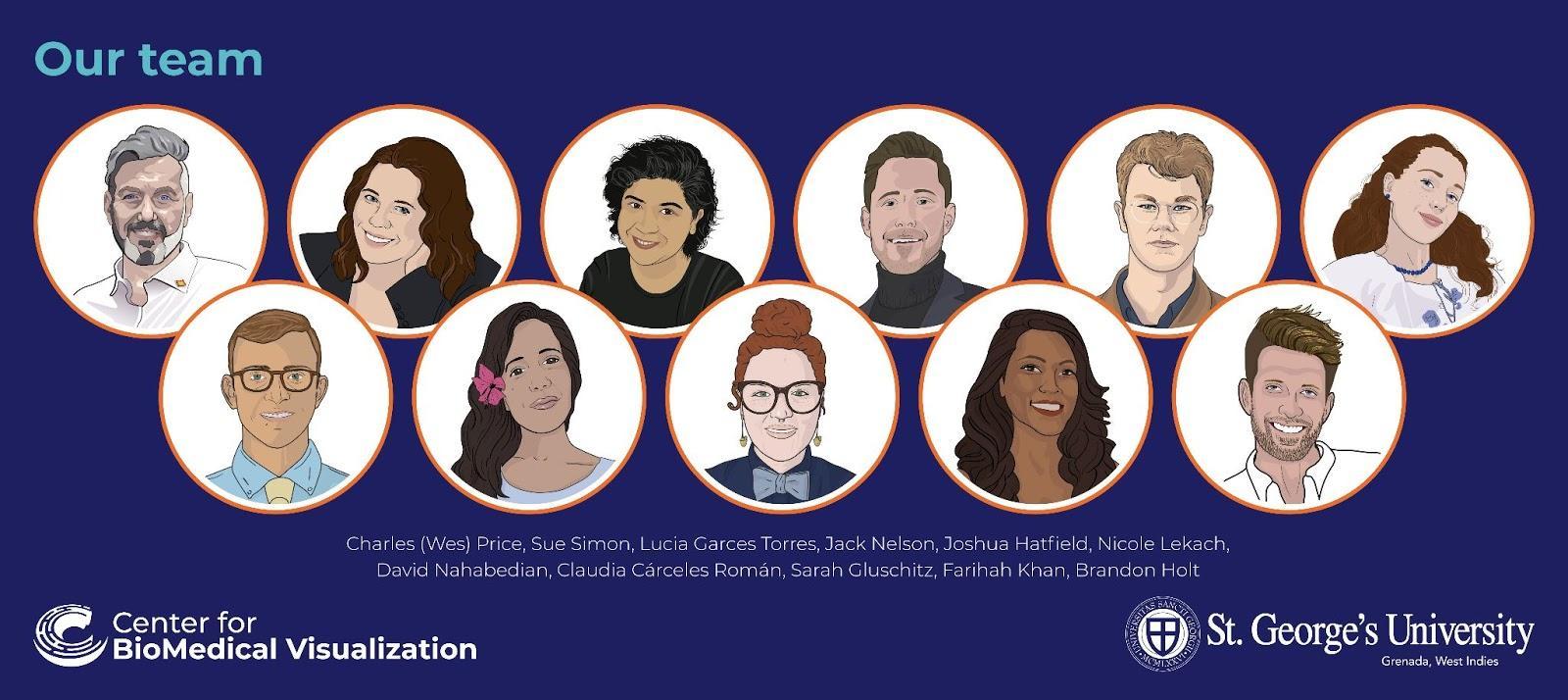
Our unit is led by Charles Wesley Price MS, CMI, and comprises faculty from all around the world:
Training in 3D
In addition to teaching anatomy to medical students, each of our team members have trained in 3D applications and software, recognizing the inherent value of 3D for the nature of our work. The 3D capabilities and backgrounds for our group range from beginner to advanced members that have been practicing in the field for years. The varied and unique skill sets, coupled with international training, has created a melting pot of talented artists and designers who share ideas, software tips, and hold internal 3D continuing education classes for our group. We actively practice, research and teach 3D modeling, 3D printing, photogrammetry, 3D scanning, animation, and 3D model extraction from DICOM data (standard file format for medical imaging such as MRI’s or CT scans).
Here is an example from our faculty that merges methods of 3D scanning and 3D modeling, created by Farihah Khan and David Nahabedian:
Why we use 3D
The initial vision for creating such a unique unit at St. George’s University was started by the renowned anatomist and scientific author, Dr. Marios Loukas, the Dean of the School of Medicine at St. George’s University. He saw the value in having faculty within the Department of Anatomical Sciences that in addition to their teaching responsibilities can develop innovative, customized, and engaging educational tools for the medical students.
Medical illustrators and visual communicators have always had an affinity for using the newest forms of technology to push the boundaries of comprehension. Traditionally, medical illustrators would use techniques such as pen and ink to draw 2D images of 3D anatomical structures. With the advancements of 3D visualization techniques, our faculty in the unit of the Center for BioMedical Visualization are creating anatomically accurate models that can be rendered from any angle, or let our audience interact with the model itself in a live viewer such as Sketchfab. Anatomy in any regard— whether it is human, veterinary, or even at the cellular level— is exceptionally difficult to
conceptualize. Learning these concepts from only 2D forms is successful, but the leap in comprehension when these models are shown in three dimensions is undeniable. Today the internet as a whole, including journals and other platforms, is jumping into the mix and making these models more accessible and easier to share with our audiences.
One of my favorite models from our team demonstrates why medical and scientific illustrators are using 3D more and more. It is The Tour of the Cell by Sarah Gluschitz, MA. It highlights the basic anatomy of a cell, takes the viewer on a ‘tour’ of a eukaryote, and shows the structures in 3D space:
Our toolkit
Our team uses a number of methods to 3D capture or build models for our educational materials, including: Photogrammetry, 3D scanning, MRI/CT extraction, 3D modeling and printing. Some of the more common software throughout the office that we use to create, process, or optimize our models and scans are: Pixologic ZBrush™, Autodesk Maya™, Maxon Cinema 4D™, and Artec Studio Pro™.
3D Scanning: 3D Scanning with the Artec Space Spider 3D Scanner™ has really become one of our strongest and most efficient ways to 3D capture specimens and anatomy for our team. We currently use the Artec Laser Scanner™ for all of our 3D scanning. Our team created a simple setup in a space that we use to scan small specimens and objects. With basic office lighting and a turntable, we can create a high fidelity scan in a matter of minutes and process the scan data, using Artec Studio Pro 13™.
Another attribute of the scanner that we take advantage of is its portability. With only needing to transport a high-powered laptop and the scanner in its protective case, we can easily take the scanner into a lab setting or other venue and start scanning within a matter of minutes. Some specimens are held in a veterinary or human cadaver lab, so transporting the specimen to the scanner isn’t always an option. Below is an example of the quality anatomical scans we are capable of creating (scan by Emily Kearney-Williams):
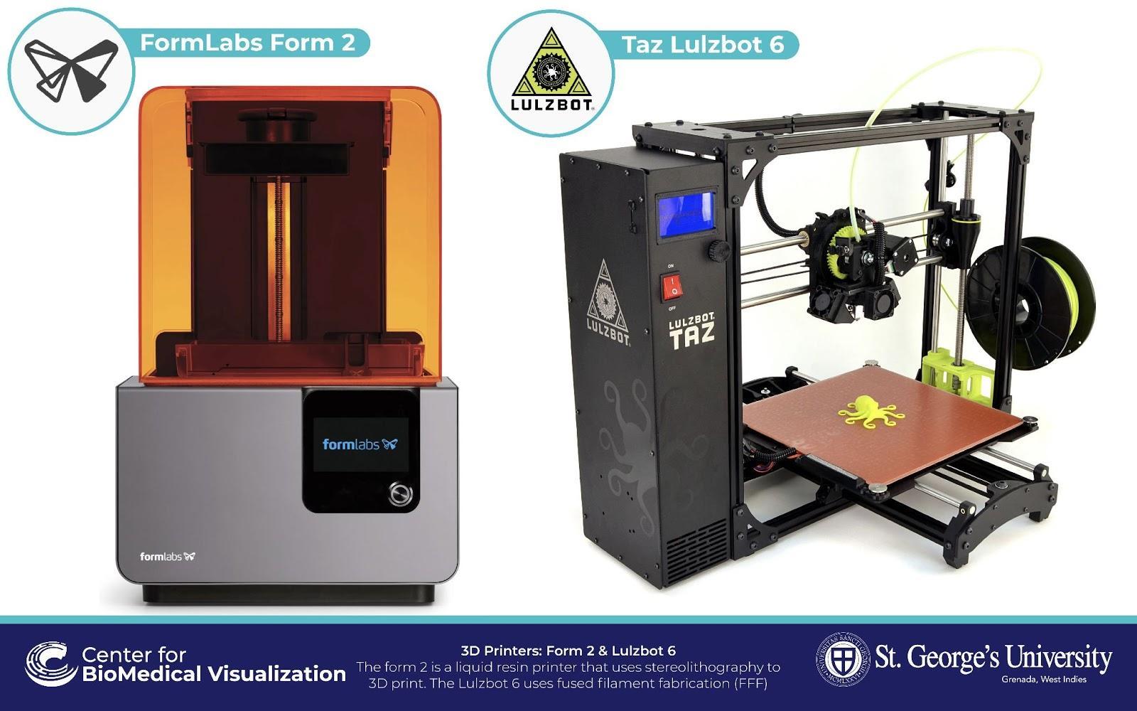
3D Printing: The Center for BioMedical Visualization is also exploring the use of 3D printing for educational purposes for St. George’s University. We currently use two forms of printing on two different printers. Our specialty printer is a FormLabs Form 2™ that uses resin-based stereolithography (SLA). This is a type of 3D printing technology that produces a print in a layer-by-layer fashion using photopolymerization, a process by which light causes chains of molecules to link, forming polymers. Those polymers then make up the body of a three-dimensional solid. Our second printer is a Taz Lulzbot™ that uses a 3D printing process that extrudes a continuous filament of a thermoplastic material, fed from a large coil through a moving, heated printer extruder head. Molten material is forced out of the print head’s nozzle and is deposited on the growing workpiece. This can also be called fused deposition modeling (FDM)™.
Software: Pixologic ZBrush™, Autodesk Maya™, and Maxon Cinema 4D™ are our team’s main tools for modeling, texturing, processing, and animating our models. Each piece of software offers different tools and methods for us to work with, which we actively use to create our Sketchfab models and animations. 3D modeling software is vital in creating anatomical 3D models that cannot be scanned or captured from life. Shown below is an example of a 3D model created by Jack Nelson BA in Maxon Cinema 4D that shows the embryological development of the heart, a concept that has always been difficult for students to visualize from 2D resources:
Below are screenshots from our team actively designing our educational Sketchfab models in their respective software (1. Jack Nelson BFA using Maxon Cinema 4D, 2. Brandon Holt MS CMI designing in Maya, 3. David Nahabedian MS CMI using ZBrush):
Photogrammetry: We have successfully created a number of photogrammetry 3D models and given professional presentations at conferences about our methods and levels of success. We have even utilized Sketchfab at these presentations so our audience members could directly interact with our models.
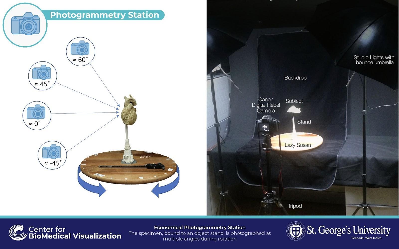
A great advantage to photogrammetry is that it is an economical and accessible way to capture and scan real-life objects and get excellent results. We use a simple setup with a digital camera and turntable. We also have written a simple guide to an economical and easy way to create your own photogrammetry setup and models at your home or office. We used the setup shown above to capture one of our cardiac plastinated specimens from the cadaver lab at St. George’s University. Photos were taken from multiple angles on a turntable to produce a high-quality and economical model:
Sketchfab & The Center for BioMedical Visualization
Sketchfab fits into our team’s workflow in a number of ways and has been vital in how we share our 3D models with clients, students and researchers. Being able to just send a link or embed a 3D model has eliminated the need for our own 3D viewing program or custom programming. Our team can really focus on making great art and sharing it without having to learn a new skill set or build a platform to share our work.
Sketchfab is also a great learning tool and we use it constantly in our internal continuing education to demonstrate model optimization and map usage to our team’s less experienced or new 3D artists. We also all browse Sketchfab to get inspired! The number of talented artists and designers using Sketchfab to showcase their work always inspires our team and challenges us to push ourselves to the next level. Another attribute about Sketchfab that we appreciate is the presentation of our models and the Center as a whole…on any device and from anywhere in the world. We have a single place to showcase our work, build collections of models and get our audiences to interact with a high-quality model within seconds of sending a link to them.
Conclusion
People experience the world in 3D. Now that we have access to these technologies, science organizations are taking advantage of them more than ever before. 3D visualization allows for a certain level of comprehension that you otherwise wouldn’t get in the traditional 2D world. These tools that have populated the 3D world in the past few years are not just flashy and cool, they are practical, innovative, and engaging. 3D modeling, scanning, and printing have changed everything.


