About
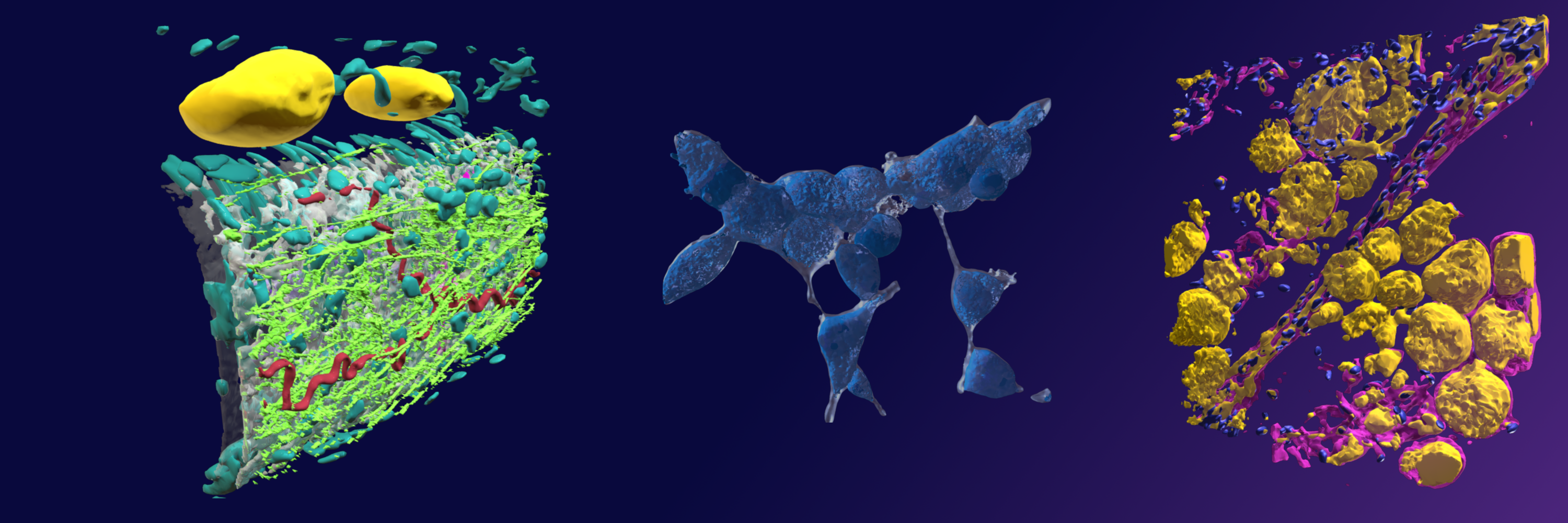
I’m Annabel Slater, a researcher at the School of Life Sciences, University of Glasgow in Scotland. Together with Dr. Craig Daly, I transform microscope images into 3D models. Our collection ‘Glasgow Life Sciences’ showcases a new use of microscopy data.
I’ve always been fascinated by the shape and form of living things. I also like how new media presents original opportunities to communicate science. This led me to take an MSc in Medical Visualisation at the University of Glasgow. Traditional science illustration consists of artists creating representations of real phenomena. Beautiful, but there’s a wealth of real microscope imagery out there that most people don’t get to see. With modern digital technology, we can transform these images into fully rendered 3D models that can be used in education, research, mixed reality apps, and animation.
Typical Workflow
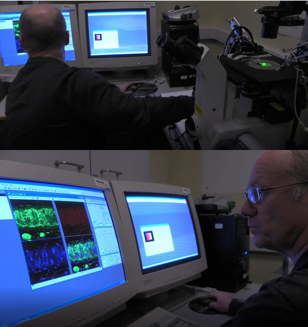
It’s important that our models are accurate representations of real scientific data produced by researchers of the University of Glasgow. We mainly work with confocal laser microscopy images. In this type of microscopy, a laser beam is focused on a biological sample in which the different components have been chemically labeled. The labels fluoresce under the light beam, and the fluorescence is captured as a series of images by the microscope.
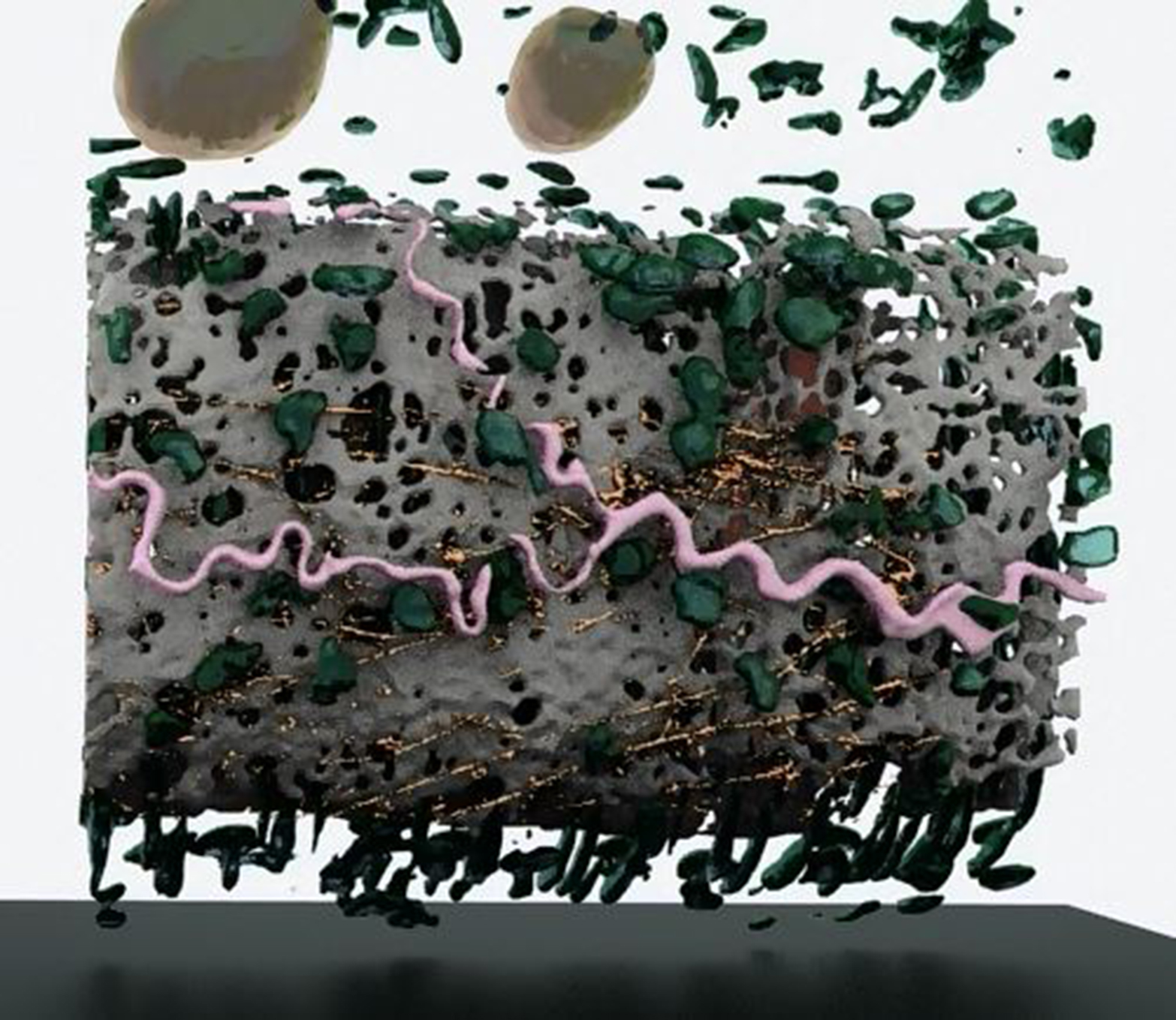
I use 3D Slicer to section out the different channels of fluorescence, then export the data in a 3D file format. The file is usually huge due to the high level of detail in this microscopy, so I use mesh reduction and retopology software to bring it down to a more manageable level. I can further refine the appearance or complexity of the model using a 3D modeling program such as 3ds Max—I might crop out sections, or remove fragments. Currently, I make use of Sketchfab’s useful suite of editorial tools to add colour and light effects to the models—it’s really easy to try out different effects.
Challenges
One difficulty is that there’s a wealth of different software available. Another difficulty is reducing the mesh without losing too much resolution. Handling large files results in long processing times. Also, it’s important to work with the researcher who provided the data in order to add additional information and confirm it looks okay—only a specialist will know whether the segmentation has captured the right elements.
The Future
Every day, 3D software and virtual reality technology are becoming more affordable and widespread. Sketchfab provides a platform to easily show and share science visualisation. Most importantly, it creates a platform to share and revive microscopy data that would otherwise languish on forgotten disk drives or in paywalled journal articles. We foresee a future in which researchers can enhance collaboration by sharing 3D models in mixed reality, and educators can teach classes using virtual reality apps containing realistic models. Students could log onto Sketchfab to view cellular models just using cardboard VR viewers, or build their own learning applications.
360° view of GLS gallery:
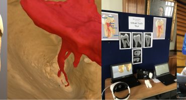
We’ve already created a virtual reality gallery to showcase our collection. The response at events has always been fantastic—people love how interactive scientific models become in virtual reality.
Short video of GLS gallery:
We plan to put the gallery online for download, and to encourage others to use our collection to build their own. It’s time for a virtual reality revolution in higher education—a new way of exploring life science.
Find us on:


