About my work at the Melbourne Dental School
I am a teaching and research academic at the Melbourne Dental School. I teach human anatomy, and my research is in mineralised tissues (bone and teeth). As part of my work studying mineralised tissues, I began learning about how to use the dental school’s microCT scanner to scan teeth and very small animal skulls—including how to turn scans of skulls and teeth into virtual models. I eventually went to Belgium to learn how to use the scanner at the Bruker factory.
My worlds collided when my love of anatomical collections (particularly teeth and skulls, both animal and human) met my ability to scan them and turn them into virtual models and 3D prints. I wanted to use these tools in teaching and learning, but also to benefit scientific researchers and students everywhere.
The versatility of virtual models in terms of what you can see, and how you can manipulate them added to my passion. I loved the fact that I could scan a skull using CT, and then use software to shave away parts of the bones virtually to reveal how the teeth are embedded in the jaws, or zoom right in to show details in the teeth that aren’t obvious to the naked eye, like a section of mouse mandible that’s smaller than a grain of rice in real life.
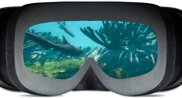
It’s truly exciting to be one of the first people to look at a newly scanned skull and fly in through one of the openings to look at the features on the inside—something no-one else has yet seen.
Some of our virtual models have been used on the cover of textbooks too, because they look so clear and crisp (The Teeth of Mammalian Vertebrates and The Teeth of Non-Mammalian Vertebrates).
My first link between virtual models and 3D printing and education came with an Engagement grant from the University of Melbourne, to engage dental students with 3D models and printing. Some of the early models on our Sketchfab account came directly from that project:
Workflow
Typically, for the projects I’m involved in, CT or microCT scanning is the preferred method over SLS or other forms of scanning. This is because we’re interested in both the external and internal structure of teeth and skulls.
-

- Loading a tooth in the microCT chamber for scanning.
-
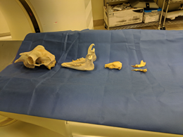
- A line-up of skulls on a clinical CT scanning bed.
Both clinical CT and microCT (just a much smaller, higher resolution version of clinical CT) are used in our projects to create virtual models.
The CT image stacks are then loaded onto 3D Slicer. We use thresholding to highlight the mineralised tissue, and then use the software to create an .stl file.

Depending on the quality and resolution of the CT scans, I use Meshlab to clean and smooth the surfaces.
It can take hours to scan skulls and teeth at the larger end of what the microCT scanner can hold. The largest object would be roughly the size of a hen’s egg (an example is the marsupial mouse skull which is a few centimetres long, and took six hours to scan). Anything larger than that requires the use of a CT scanner.
The human teeth I scan take about one to one and a half hours in the microCT scanner.
Scan files are usually very large for CT and microCT, and one of our biggest challenges was having the computing power and storage capacity to handle all of our data. This has changed for the better significantly over the last decade, I think because of the increase in gaming technology and requirements!
Our virtual models on Sketchfab
Two years ago, a group of colleagues and I were successful in getting a grant to scan some skulls and teeth from different scientific collections across the University of Melbourne. We started adding these models to the existing Sketchfab page. We did that because Sketchfab enables us to reach a much greater audience for these special scans. Just as the skulls are diverse and interesting, you never know who the models will help in the big world outside the University: school students, researchers, conservationists, archaeologists, and dental professionals…the list goes on. We wanted to make the models as accessible as possible, and Sketchfab helps us to do that.
Sketchfab made it easy to tailor the display of our models in terms of lighting and texture. It also has ‘like’ and comment functions. I really enjoy interactions with people centred around the models. It’s like social media for virtual model enthusiasts. The share function is also very useful for us when we want to show examples to collaborators, colleagues, and students.
The future of 3D science
I think we will move further towards not just scanning and recreating structures virtually, but then creating something virtually and bringing it into the real world. Teeth are the perfect example, and if you think about the way your body makes teeth, it’s already like additive manufacturing.
The versatility of 3D models (both printed and virtual) means that researchers and students can have better access to these resources. A lot of things that could only be done once on a skull—cutting it in half to see the inside, for example—are now possible in infinite ways without destroying the original. Being able to do it over and over again allows us to learn a lot of different things, and do lots of measurements and calculations, etc. using virtual scans. This approach increases sample sizes for researchers, and also supports animal ethics and welfare, because the skeletal material can be reused.
I think our virtual world will expand more and more, and that will help to support our real-life world, especially for health and learning. It’s an exciting time, and all we have to do is let this technology keep capturing our imaginations!
About the author
Rita Hardiman
I am a senior lecturer and researcher in anatomy and mineralised tissues at the Melbourne Dental School, part of the University of Melbourne in Victoria, Australia. I have loved the range of possibilities that 3D scanning and printing enable for researching and learning about teeth and skulls. I have been scanning skulls and teeth for about six years, and 3D printing for five. The materials I work with are ideal for scanning and printing, and I love how the process maximises the information available from the objects. You can find me on https://findanexpert.unimelb.edu.au/profile/3920-rita-hardiman or twitter: @ritahardiman