About
My name is Lincoln Wong and I am a pediatric radiologist at Children’s Hospital and Medical Center in Omaha, Nebraska. As a radiologist, I am a physician who uses imaging to help diagnose disease. These images can be made from x-rays, ultrasounds, MRI or CT scans, traditionally viewed as 2-dimensional images on a computer screen. In the last 3 years, Children’s has embarked upon finding new ways of presenting this data, converting 2D images into 3D models for AR, VR and 3D printing. Sketchfab has been a critical tool for making this happen.
3D at Children’s Hospital and Medical Center
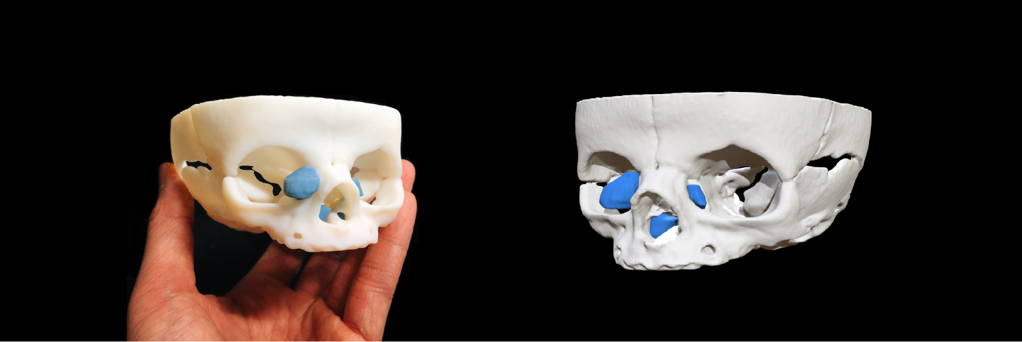
3D imaging brings patient cases to life. Complementing my written report, a 3D model can communicate many findings more efficiently than the written report on its own. A surgeon can look at the 3D model of a patient and know very quickly what the original scan depicted or what my report described. For a surgeon who thinks and operates in a 3D world, a 3D model allows them to see very easily complex disease processes that would otherwise be difficult to read in a written report. These models give surgeons added confidence that they know precisely what lays ahead prior to surgery. Reviewing a model can be a decisive step in surgical planning.
Furthermore, models are used to educate medical trainees as well as help patients and family members understand a complicated disease process they face. Without a medical background, showing them traditional radiology images can be very abstract; showing them a 3D model elevates their understanding and may ease some anxiety as they go through this stressful period.
We have used 3D imaging for a variety of cases. Surgical oncology, craniofacial surgery, cardiothoracic surgery, neurosurgery, ophthalmology, urology, orthopedics, otolaryngology, pulmonology and gastroenterology are counted among the medical specialties we help with 3D imaging.
Workflow
Our workflow begins by exporting and anonymizing a patient’s CT and MR data. Using segmentation software Materialise Mimics Medical, the images are converted into 3D STL files by tracing out each desired anatomical part. This can be done with the help of computer-generated segmentation or manual segmentation. I then use Blender to edit the model and prepare it for uploading on to Sketchfab.
On Sketchfab the model is easy to share and view with the care team, medical trainees, patients and families. I can send surgeons a link that they can use to easily review the case on their smartphone, tablet or computer. We use Sketchfab to preview our cases and to decide if we want to take the model further into AR, VR, or 3D printing.
Looking Ahead
Finding enough time in the day to oversee the many projects we have in our 3D Printing and Advanced Visualization lab can be challenging. However, the gratitude I get from our providers and knowing that I’m helping a child receive the best possible care with positive outcomes pushes me to drive on. I am also lucky enough that this endeavor blends my passions for art and science.
One of my favorite stories that came out of our lab is helping to produce and print a 3D model of the face of a fetus for a blind mother. The 3D ultrasound images were converted into a 3D file which was subsequently printed on our 3D printer and presented to the mother. Cases like this make one realize how powerful this technology can be.
3D visualization in medicine is in its infancy with the potential to transform healthcare, medical education, and elevate the patient experience. We are still finding new applications for this technology. The Radiology Society of North America has formed special interest groups who meet and share ideas on 3D printing, AR and VR applications. It is exciting to be at the ground level and help shape where this technology will go and discover new ways that we can provide better care for our patients.


